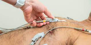
ECHO-CARDIOGRAM

An echocardiogram, also known as an echo, is a type of imaging test that uses sound waves to create images of the heart. It is a painless and non-invasive procedure that is often used to:
- Diagnose heart problems, such as heart valve disease, heart failure, and congenital heart defects
- Assess the size, shape, and function of the heart
- Monitor the effectiveness of heart treatments
An echocardiogram is typically performed by a trained technician called a sonographer. During the test, you will lie on your back on an examination table. The sonographer will place a transducer, which is a small, hand-held device, on your chest. The transducer emits sound waves that travel through your chest and bounce off your heart. The reflected sound waves are then converted into images of your heart that are displayed on a video monitor.
There are different types of echocardiograms, each of which provides different information about the heart. The most common type of echocardiogram is a transthoracic echocardiogram, which is performed by placing the transducer on your chest wall. Other types of echocardiograms include:
- Transesophageal echocardiogram: This type of echo is performed by placing the transducer in the esophagus, the tube that connects your mouth to your stomach. This provides a closer look at the heart, which can be helpful for diagnosing certain conditions.
- Stress echocardiogram: This type of echo is performed while you are exercising or taking medication that makes your heart beat faster. This can help to identify blockages in the heart arteries.
- Fetal echocardiogram: This type of echo is performed on pregnant women to check the health of the baby's heart.


